
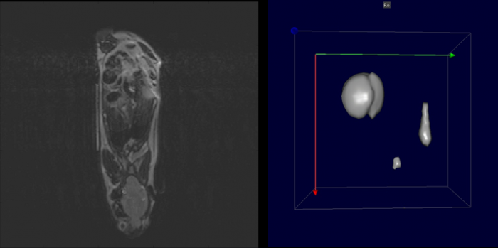
 Left: MRI slice showing superior view of mouse anatomy with marker capillary filled with SPIO marker. Right: SPIO marker as seen in 3D by the MPI system.
Reconstruction and registration of images acquired by multimodal preclinical imaging methods
Left: MRI slice showing superior view of mouse anatomy with marker capillary filled with SPIO marker. Right: SPIO marker as seen in 3D by the MPI system.
Novel functional imaging modality requiring co-registration with anatomical information for in vivo imaging.
We are investigating the use of a novel functional imaging modality requiring co-registration with anatomical information for in vivo imaging. This project is done in cooperation with Center for Advanced Preclinical Imaging (CAPI), First Faculty of Medicine (Charles University, Prague), in the framework of Czech-BioImaging project (https://www.czech-bioimaging.cz/).
Magnetic particle imaging (MPI) makes use of magnetic nanoparticles and high intensity magnetic fields for high-resolution, high-speed functional imaging. In the small animal studies conducted in this project, MPI data must be combined with other preclinical imaging methods such as magnetic resonance imaging (MRI) and computed tomography (CT) for useful information.
Obtained datasets are co-registered using P-MOD (PMOD Technologies, Germany) and MATLAB (MathWorks, USA) to obtain 2D and 3D visualizations of the contrast agents in the tissue. Automatic and manual methods of co-registration will be employed until a suitable protocol is developed. Examples of the set-up and preliminary results are shown above.
For more information, please contact Kevin Loo, PhD (kevin.loo@fgu.cas.cz) or visit CAPI’s website.