TMEM70 disease
Animal models of human mitochondrial diseases enable detailed insight into pathogenic mechanisms, from molecular to organismal levels.
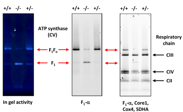
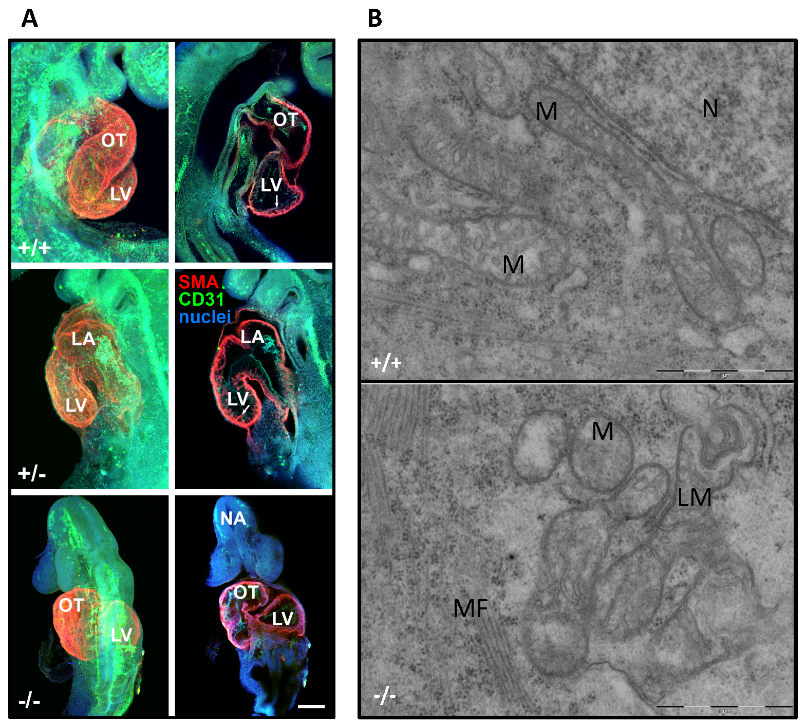
According to previous studies performed at the Institute Physiology CAS, the mitochondrial ATP synthase, the key producer of cellular ATP, uniquely depends on a TMEM70, a 21-kDa protein that facilitates the biogenesis of mammalian enzyme. TMEM70 gene mutations turned out to represent the most frequent cause of ATP synthase deficiency resulting in a severe mitochondrial disease, neonatal encephalo-cardiomyopathy in humans (OMIM 604273). To further study this ancillary factor in vivo, Tmem70-deficient mice was generated. Tmem70 ablation resulted in profound growth retardation and embryonic lethality at 9.5 days of fetal development. Affected embryos displayed 80% decrease of ATP synthase and impaired biogenesis of ATP synthase, stalled at the early stage, following the formation of the F1 catalytic part of enzyme. Consequently, a decrease in ADP-stimulated respiration, respiratory control and ATP/ADP ratios, demonstrated compromised mitochondrial ATP production. Tmem70-/- embryos exhibited delayed development of the cardiovascular system and disturbed heart mitochondrial ultrastructure. These results thus showed that Tmem70 knockout in the mouse leads to embryonic lethality due to the lack of ATP synthase and impairment of mitochondrial energy provision and fully confirmed the expected mechanism of human TMEM70 disease. Using an inducible Tmem70 knockout, which allows us to study the function of the Tmem70 protein in living mice, we further found that a deficiency of the Tmem70 protein leads to the assembly of incomplete ATP synthase complex (cV*), which lacks the so-called c-ring. This finding represents the first clear evidence that the main role of Tmem70 is to assist in the assembly of the c-ring, a key structure for ATP synthase function.


ATP synthase deficiency and accumulation of F1 subcomplex in Tmem70-/- embryos.
Blue-Native electrophoresis of ATP synthase (F1Fo) and respiratory chain complexes of wt (+/+) null (-/-) and heterozygous (+/-) Tmem70 knockout E9 embryos. ATPase in gel activity and WB detection with antibodies to ATP synthase (F1-α) and respiratory complexes II-IV (CII – SDHA subunit; CIII – Core 1 subunit; CIV – Cox4 subunit).

Impaired morphology of Tmem70-/- embryos.
(A) Confocal microscopy of retarded heart in null (-/-) embryo in comparison with wt (+/+) and heterozygous (+/-) E9.5 embryos. OT – outer tract, LV – left ventricle, LA – left atrium, NA – neuroporus anterior. Immunohistochemistry: SMA (alpha smooth muscle actin), red – myocardium; CD31, green – endocardium; Hoechst 33342, blue – nuclei; arrows – emerging ventricular trabeculation, scale bar 100 µm. (B) Electron microscopy of disturbed cristae morphology in null (-/-) compared to wt (+/+) heart mitochondria of E9.5 embryos. M – mitochondria, MF – myofibrils, LM – lysed mitochondria, N – nucleus, scale bar 1 µm.
Vrbacký, Marek – Kovalčíková, Jana – Chawengsaksophak, Kallayanee – Beck, Inken – Mráček, Tomáš – Nůsková, Hana – Sedmera, David – Papoušek, František – Kolář, František – Sobol, Margarita – Hozák, Pavel – Sedlacek, Radislav – Houštěk, Josef. Knockout of Tmem70 alters biogenesis of ATP synthase and leads to embryonal lethality in mice. Human Molecular Genetics, Vol. 25, No 21 (2016), p. 4674-4685. IF=5.985
Composition of incomplete ATP synthase (cV*)

Native electrophoresis of ATP synthase in mouse liver homogenate; control (+/+) vs. inducible Tmem70 knockout (-/-). Act – detection of ATP synthase activity in gel; F1-α, Fo-a, Fo-b, Fo-c – detection of ATP synthase subunits using specific antibodies.
Kovalčíková, Jana – Vrbacký, Marek – Pecina, Petr – Tauchmannová, Kateřina – Nůsková, Hana – Kaplanová, Vilma – Brázdová, Andrea – Alán, Lukáš – Eliáš, Jan – Čunátová, Kristýna – Kořínek, Vladimír – Sedláček, Radislav – Mráček, Tomáš – Houštěk, Josef. TMEM70 facilitates biogenesis of mammalian ATP synthase by promoting subunit c incorporation into the rotor structure of the enzyme. FASEB Journal. 2019; 33(12); 14103-14117. IF = 4.966
