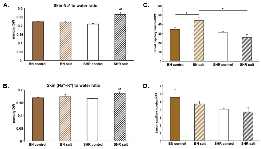
Recent studies in humans and rats have suggested that increased Na+ deposition in the skin without concomitant water retention may predispose to salt-sensitive hypertension. Therefore, we compared tissue Na+ deposition in salt-sensitive spontaneously hypertensive rats (SHR) versus salt-resistant normotensive Brown Norway (BN) rats. After salt administration (10 days of drinking a 1% NaCl solution), SHR rats showed a significant parallel increase in both the Na+-to-water and (Na++K+)-to-water ratios, suggesting increased deposition of osmotically inactive Na+ in the skin, whereas no significant changes in skin electrolyte concentrations were observed in BN rats. In SHR rats, there was a non-significant decrease in the number of skin blood capillaries (rarefaction) after salt administration, whereas in BN rats, there was a significant increase in the density of blood capillaries in the skin. Analysis of gene expression profiles in the skin of BN rats after salt administration showed a significant increase in the expression of genes involved in angiogenesis and endothelial cell proliferation, in contrast to SHR rats. Given that the majority of the body’s resistance vessels are located in the skin, it is possible that capillary rarefaction may lead to increased peripheral resistance and salt sensitivity in SHR rats (Šilhavy et al, 2022).

Electrolyte concentrations in the skin of SHR and BN strains. (A) Salt administration was associated with a significant increase in the Na+- to-water ratio (B) and also in the (Na++K+)-to-water ratio in the skin of SHR rats. (C) The number of blood capillaries in the skin of the BN strain was significantly increased after salt administration in contrast to the SHR strain. (D) No significant differences were observed in the number of lymphatic capillaries in the skin of the BN and SHR strains between the strains and the amount of salt administered.


