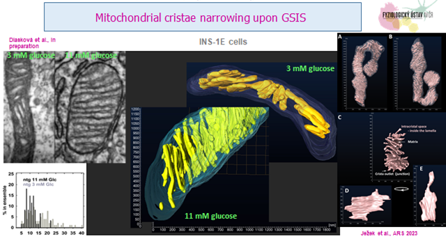
In 2018, we reported narrowing of mitochondrial cristae upon glucose-stimulated insulin secretion (GSIS) (Dlasková et al, BBA 2018 ;1859 (9):829-844), which was similar as cristae narrowing of hypoxia-adapted HepG2 cells after a sudden substrate intake (Dlasková et al, BBA 2019; 1860(8):659-678). Recently, we excluded the role of membrane FO-moiety of the ATP-synthase in shaping of cristae and in driving these cristae ultramorphology changes (v přípravě); suggesting an alternative in osmotic forces which should drive cristae inflation/narrowing (Ježek J et al, ARS 2023 ;39(10-12):635-683).
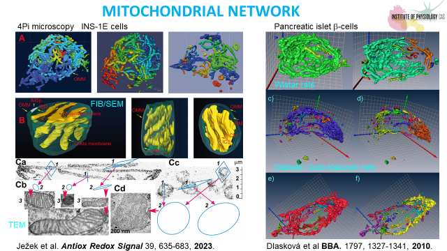
The role of other cristae shaping proteins such as various isoforms of OPA1, arising from splicing and cleavage by specific proteases; MICOS subunits and prohibitins i salso studied. Cristae investigations include classic transmission electron microscopy (TEM) and 3 dimensional microscopies such as FIB/SEM (focused-ion beam/scanning electron microscopy) and 3D dSTORM (direct stochastic optical reconstruction microscopy) which will be complemented by 2D superresolution STED microscopy.
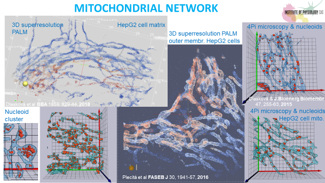
In 2023 we have completed our previous series of papers concerning nucleoids of mitochondrial DNA and using 3D dSTORM, demonstrating the existence of more than one mtDNA molecule per nucleoid at least in certain nucleoid fraction (Pavluch et al, SciRep 2023 ;13(1): 5788). We will attempt to develop electron microscopies to visualize mtDNA nucleoids.
These projects are supported by grants 24-10325S, 23-05798S and 22-02203S of the Czech Science Foundation (GAČR).


