In 2015, we revealed the role of Ca2+-independent mitochondrial phospholipase iPLA2g/PNPLA8 in fatty acid-stimulated insulin secretion (FASIS) (Ježek J et al, ARS 2015 ; 23(12):958-72). ). In 2020 we discovered the essential role of redox (H2O2) signal ensured by the NADPH-oxidase, isoform 4 (NOX4) in glucose-stimulated insulin secretion (GSIS) (Plecitá et al, Diabetes 2020 ; 69(7): 1341-1354).
We also described pyruvate-dependent redox shuttles switched upon GSIS, transforming mitochondrial NADH into the cytosolic NADPH to feed NOX4 (Plecitá et al, ARS 2020 ; 33(12):789-815). Finally, we quantified the redox signal (H2O2) upon FASIS and GSIS, distinguished metabolic and GPR40 receptor branch of FASIS and demonstrated FASIS in vivo responding to incoming chylomicrons in mice (Jabůrek et al, Redox Biol 2024 ; 75:103283).
Our current studies extend these seminal papers, investigating on branched-chain keto acid- (BCKA-) induced redox signals and insulin secretion, crosstalk between BCKA and fatty acid metabolism and determining the role of lipid droplets (LDs) in GSIS and FASIS. Redox signals are studied also using an optogenetic approach. FASIS is further studied in vivo investigating on the concept of exaggerated FASIS as participating in type 2 diabetes etiology.
Since mice with NOX4 ablated specifically in pancreatic beta cells exhibit insulin resistance, we seek for messengers of redox disequilibria in beta cells reporting such state to the periphery or neighbor acinar and ductal pancreatic cells.
These projects are supported by grants 24-10132S and 22-17173S of the Czech Science Foundation (GAČR) and by the project National Institute for Research of Metabolic and Cardiovascular Diseases (Programme EXCELES, ID Project No. LX22NPO5104) – funded by the European Union – Next Generation EU.
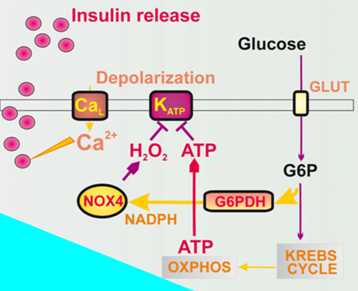
Figure 1 Redox signal from NOX4 concomitant to the elevation of ATP is essential for insulin secretion – both are required for closing of the ATP-sensitive K+channel (KATP) (from Plecitá-Hlavatá et al., Diabetes 2020, 69, 1-14).
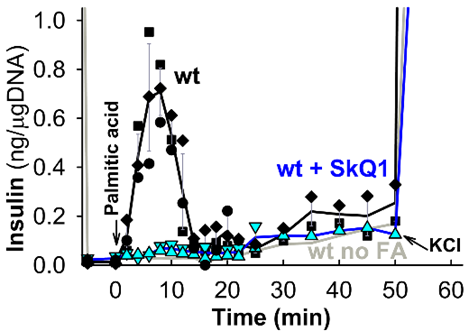
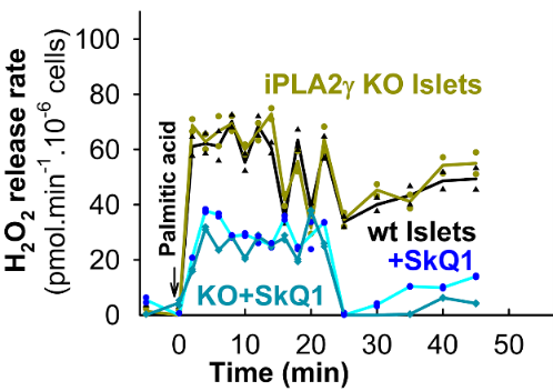
Figure 2 Perifused pancreatic islets provide insulin secretion (left) plus H2O2 release to the cell exterior (right), both prevented by mitochondria-targeted antioxidant SkQ1 (from Jabůrek et al., Redox Biol 2024; 75:103283).
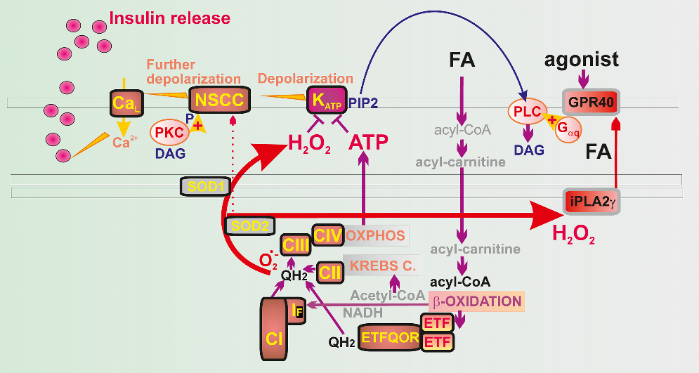
Figure 3 Redox signal from mitochondrial b-oxidation of fatty acids (FA) plus elevation of ATP are essential for FA- stimulated insulin secretion (FASIS). This redox signal (H2O2) also activates iPLA2g supplying mitochondrial FAs for GPR40 receptor pathway, removing PIP2 from the ATP-sensitive K+channel (KATP), thus withdrawing its state of a permanent closure (from Jabůrek et al., Redox Biol 2024; 75:103283).


