The modern field of biomaterials and tissue engineering is successfully developing in Institute of Physiology. A synthetic materials are increasingly used for replacements of damaged tissues and organs.
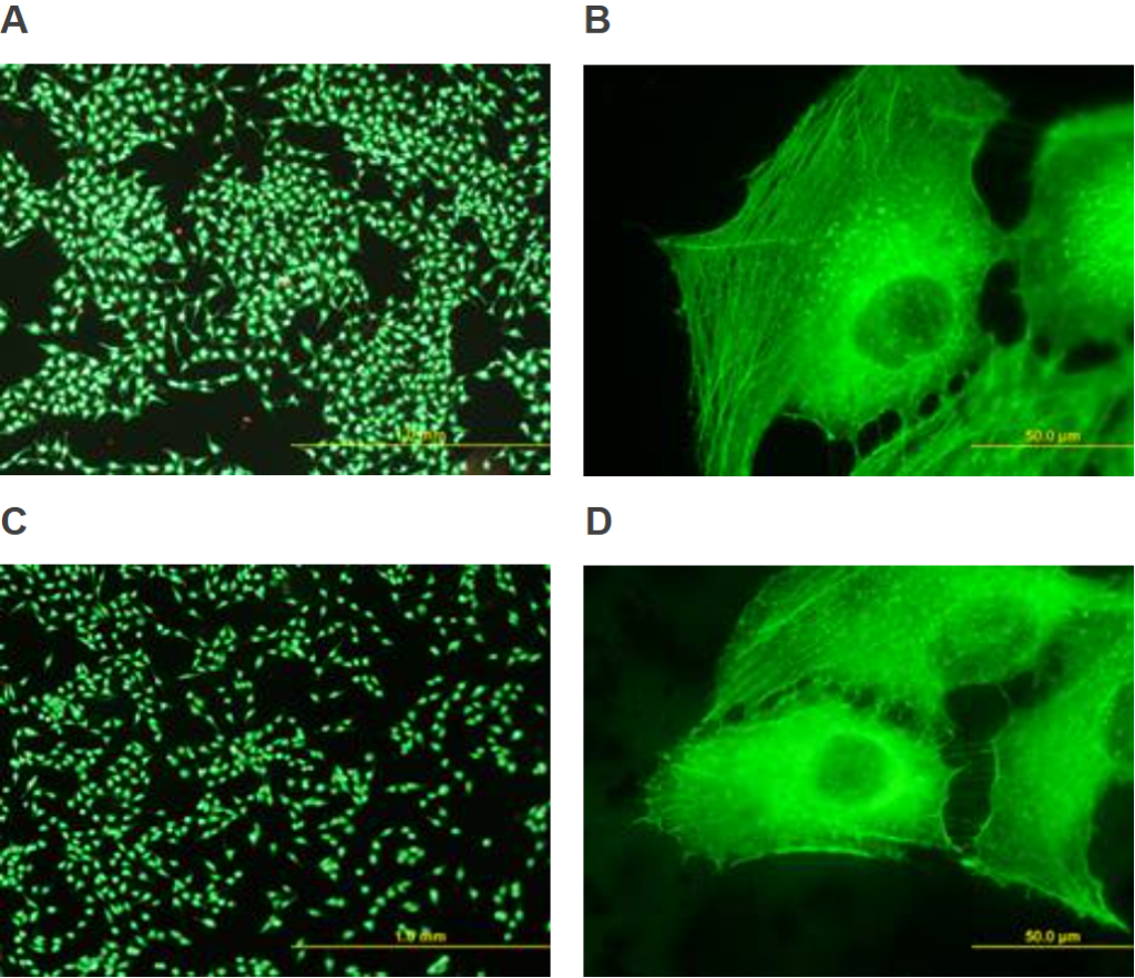
A new progressive interdisciplinary discipline of tissue engineering is developing, which uses the knowledge and methods of engineering and biological sciences. It aims to construct replacements in which the artificial material plays the role of a natural extracellular matrix and controls cell function to regenerate damaged tissue. The modern field of biomaterials and tissue engineering is also successfully developing at the Institute of Physiology. In collaboration with AGH University of Science and Technology in Krakow, we have used polysulfone reinforced with carbon nanotubes as a material component of potential replacements for damaged bone tissue. These nanotubes both improved the mechanical properties of the material and promoted adhesion, spreading, viability and growth of bone cells (Figure 1). The surface nanostructure of the material caused by the protruding nanotubes probably played a role in this, but the electrical conductivity of the nanotubes may also have been beneficial. It is known that electrically conductive materials promote cell adhesion, growth, differentiation and other cellular manifestations even without active electrical stimulation, although the passage of an electric current through the material would probably further enhance the cellular functions we observed.
Fig. 1. Human bone cells of the MG-63 line in three-day cultures on polysulfone films enriched with single-walled carbon nanotubes as nanorods (A, B) or multi-walled carbon nanotubes (C, D), both at a concentration of 2 weight %. After staining with the commercially available LIVE/DEAD kit (A, C), virtually all cells show green fluorescence, i.e. they have high viability as manifested by esterase enzyme activity. Only occasionally we find red stained cells, i.e. dead cells with damaged cell membrane. Cells on both materials are well distributed and have a well-developed fibrous beta-actin cytoskeleton (B, D). The scale bar is 1 mm (A, C) or 50 μm (B, D).
An important success of Institute of Physiology AS CR in 2014 – Dr. Bačáková and her team
Human osteoblast-like MG 63 cells on polysulfone modified with carbon nanotubes or carbon nanohorns
By: Stankova, Lubica; Fraczek-Szczypta, Aneta; Blazewicz, Marta; Filova, Elena; Blazewicz, Stanislaw; Lisa, Vera; Bacakova, Lucie
CARBON Volume: 67 Pages: 578-591 Published: FEB 2014
IF = 5.868


