Cyclin-dependent protein kinase 16 (CDK16) is an enzyme that transfers phosphate groups onto other proteins, thereby controlling their activity. In human cells, it helps regulate cell growth and survival, particularly in the brain and testes, where it plays a key role in proper sperm development. CDK16 has also been found to be frequently overactive in cancer cells, where it promotes cell division, resistance to cell death, and metastasis. A new study published in Nature Communications has revealed, at the atomic level, the regulatory mechanism that activates CDK16. A detailed understanding of CDK16 regulation is of significant biomedical importance, for example, in oncology, where it may support the development of novel anticancer kinase inhibitors.
Cyclin-dependent protein kinase 16 (CDK16) regulates a variety of physiological and pathological processes, including autophagy, spermatogenesis, and tumour growth. Unlike most related kinases within the CDK family, CDK16 is not activated solely by a cyclin but by a complex composed of phosphorylated cyclin Y (CCNY) and the adaptor protein 14‑3‑3. Until now, however, it remained unclear how exactly this trio of proteins interacts and how these interactions “switch” CDK16 from an inactive to an active state.
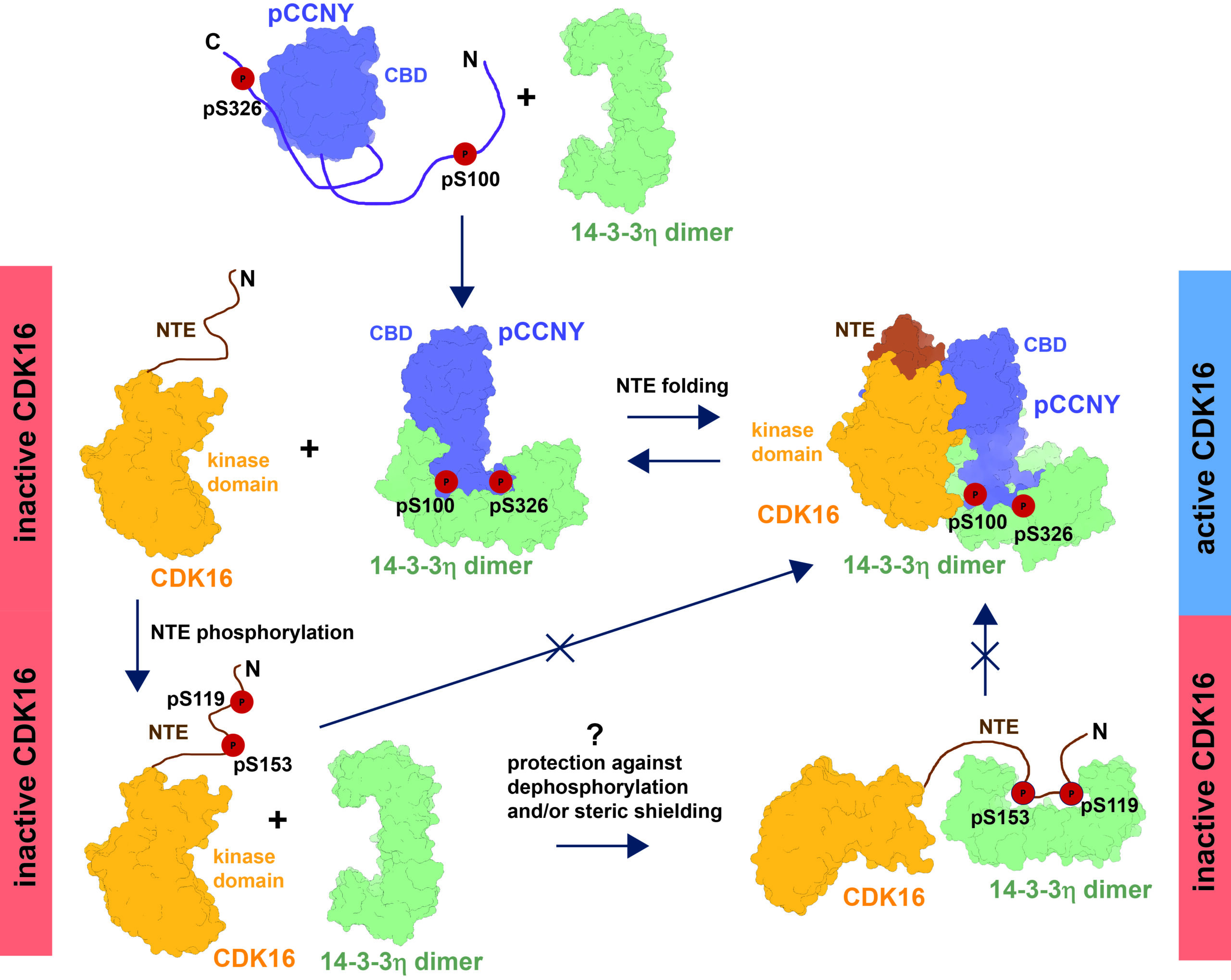
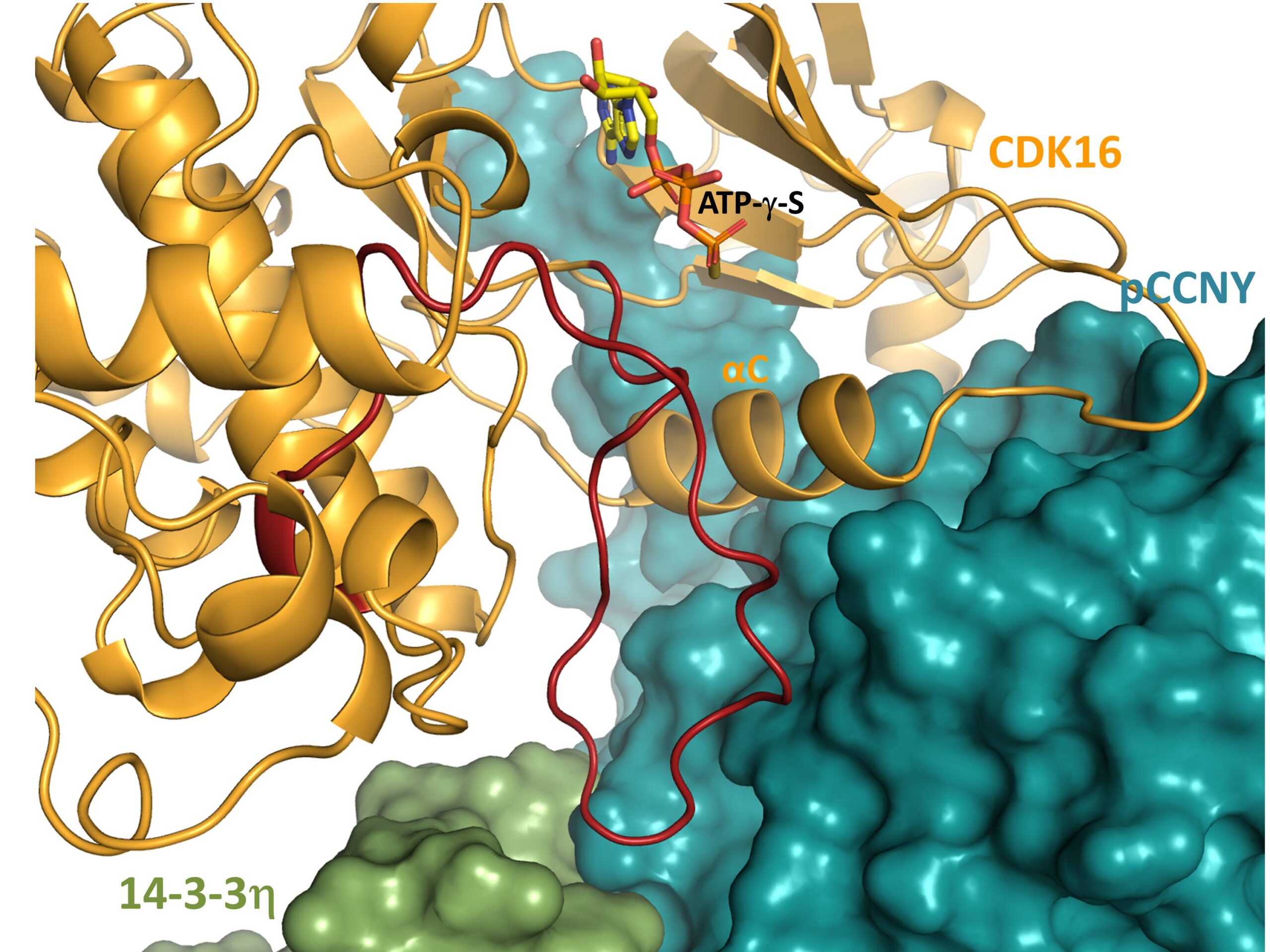
The teams led by Tomáš Obšil from the Department of Physical and Macromolecular Chemistry at Charles University in Prague and Veronika Obšilová from the Laboratory of Structural Biology of Signalling Proteins of IPHYS elucidated the structure of CDK16 in complex with phosphorylated CCNY and 14‑3‑3. “Using advanced structural biology techniques (cryo‑EM analysis and hydrogen–deuterium exchange coupled with mass spectrometric detection), we determined the 3D structure of the CDK16–CCNY–14‑3‑3 complex and tracked its dynamics. The obtained structure shows that binding of the 14‑3‑3 protein alters the arrangement of a key region of cyclin Y that forms the interaction interface for CDK16, thereby enabling kinase activation,” explains Tomáš Obšil. Full activation of CDK16 thus requires not only the classical kinase–cyclin contacts but also specific interactions of cyclin Y with the N‑terminal region of CDK16, together with the simultaneous binding of phosphorylated cyclin Y to 14‑3‑3.
“An important aspect is that cyclin Y must first be phosphorylated on specific amino acid residues in order to be recognised by the 14‑3‑3 proteins and form a stable complex with them, which then ‘shapes’ the proper binding surface for CDK16. This multilayered regulation ensures that CDK16 becomes activated only in the appropriate cellular context, for example, in response to the cell’s energy status or signalling pathways that control cell division and survival,” adds Veronika Obšilová.
Understanding the precise spatial organisation of interactions among CDK16, cyclin Y, and 14‑3‑3 is of fundamental importance for biomedicine. It enables rational design of small molecules that could specifically disrupt or stabilise these protein–protein contacts—and thereby selectively modulate CDK16 activity in cancer cells without significantly affecting related kinases. This work thus expands the structural foundation for developing new anti‑cancer drugs that target not only the kinase active site but also its regulatory complexes with cyclins and adaptor proteins.
Reference: Kohoutova K., Kosek D., Brzezina A., Honzejkova K., Obsilova V., and Obsil T.: Structural basis of the cyclin Y/14-3-3 protein-mediated activation of CDK16. Nat Commun (2026). IF = 15.7; DOI: 10.1038/s41467-026-70778-5